Infrastructure and technology development
CCFS links three internationally recognised concentrations of analytical geochemistry infrastructure: GEMOC’s Geochemical Analysis Unit (Macquarie University) and the associated Computing Cluster, the Centre for Microscopy, Characterisation and Analysis (UWA/Curtin) and the John de Laeter Centre of Mass Spectrometry. All are nodes for the NCRIS AuScope and Characterisation Capabilities, and have complementary instrumentation and laboratories. In addition, Curtin and UWA share a leading facility for paleomagnetic studies, and facilities for experimental mineralogy and petrology are being built up at Macquarie and Curtin.
Facilities:
CCFS/GEMOC INFRASTRUCTURE, LABORATORIES AND INSTRUMENTATION
CMCA TECHNOLOGY DEVELOPMENT AND INSTRUMENTATION
WESTERN AUSTRALIA PALAEOMAGNETIC AND ROCK-MAGNETIC FACILITY
CCFS/GEMOC INFRASTRUCTURE, LABORATORIES AND INSTRUMENTATION
CCFS links three internationally recognised concentrations of analytical geochemistry infrastructure: GEMOC’s Geochemical Analysis Unit (Macquarie University) and the associated Computing Cluster, the Centre for Microscopy, Characterisation and Analysis (UWA/Curtin) and the John de Laeter Centre of Mass Spectrometry. All are nodes for the NCRIS AuScope and Characterisation Capabilities, and have complementary instrumentation and laboratories. In addition, Curtin and UWA share a leading facility for paleomagnetic studies, and facilities for experimental mineralogy and petrology are being built up at Macquarie and Curtin.
CCFS/GEMOC INFRASTRUCTURE, LABORATORIES AND INSTRUMENTATION
The analytical instrumentation and support facilities of the Macquarie University Geochemical Analysis Unit (GAU) represent a state-of-the-art geochemical facility.
The GAU contains:
- a Cameca SX-100 electron microprobe
- a Zeiss EVO MA15 Scanning electron microscope (with Oxford Instruments Aztec Synergy EDS/EBSD)
- four Agilent quadrupole ICPMS (industry collaboration; two 7500cs; two 7700cx)
- two Nu Plasma multi-collector ICPMS (one decommissioned in June 2015)
- a Nu Plasma II multi-collector ICPMS (installed in June 2015)
- a Nu Attom high resolution single-collector sector field ICPMS
- a Thermo Finnigan Triton TIMS
- three New Wave laser microprobes (one 266 nm, two 213 nm, each fitted with large format sample cells) for the MC-ICPMS and ICPMS laboratories (industry collaboration)
- two Photon Machines Excite Excimer laser ablation systems
- a Photon Analyte G2 Excimer laser ablation system
- a Photon Machines Analyte198 Femto-second laser ablation system
- a PANalytical Axios 1kW XRF with rocker-furnace sample preparation equipment
- a LECO RC412 H2O-CO2 analyser
- an Ortec Alpha Particle counter
- a New Wave MicroMill micro-sampling apparatus
- a ThermoFisher iN10 FTIR microscope
- a Horiba LABRAM HR Evolution confocal laser Raman microscope
- selFrag electrostatic rock disaggregation facility
Clean labs and sampling facilities provide infrastructure for ICPMS, XRF and isotopic analyses of small and/or low-level samples.
Experimental petrology laboratories include four piston-cylinder presses (pressures to 4 GPa), hydrothermal apparatus, controlled atmosphere furnaces,Griggs apparatus and a multi-anvil apparatus for pressures to 27 GPa.
THE GEMOC FACILITY FOR INTEGRATED MICROANALYSIS (FIM) AND MICRO-GIS DEVELOPMENT
GEMOC is continuing to develop a unique, world-class geochemical facility, based on in situ imaging and microanalysis of trace elements andisotopic ratios in minerals, rocks and fluids. The Facility for Integrated Microanalysis now consists of four different types of analytical instrument,linked by a single sample positioning and referencing system to combine spot analysis with images of spatial variations in composition (“micro-GIS”). TheFIM has been in operation since mid-1999. Major instruments were replaced or upgraded in 2002-2004 through the $5.125 million DEST Infrastructure grantawarded to Macquarie University with the Universities of Newcastle, Sydney, Western Sydney and Wollongong as partners. Further enhancement of the facilitytook place following the award of an ARC LIEF grant in 2010 to integrate the two existing multi-collector inductively-coupled-plasma mass spectrometers(MC-ICPMS) with three new instruments: a femtosecond laser-ablation microprobe (LAM; installed in June 2012); a high-sensitivity magnetic-sector Nu AttomICPMS (installed in January 2013); an Agilent 7700 quadrupole ICPMS (installed in 2010). In 2012 GEMOC was awarded ARC LIEF funding for a second generationMC-ICPMS and a Nu Plasma II was installed in June 2015.
Equipment for high-pressure experimentation
The expansion of the high-pressure experimental facilities continued in 2015, with plans for an extension to the laboratory in a new wing extending intothe yard behind building E5A. This will house two large multi-anvil presses, and a third will be installed in the current laboratory. Laser-heated diamondanvil cells and an additional piston-cylinder apparatus will be acquired in 2016 through LIEF funds from the Australian Research Council. An experimentalprogram on electrical conductivity in mantle materials has begun with the currently available multi-anvil apparatus.
PROGRESS IN 2015:
1. Facility for Integrated Microanalysis
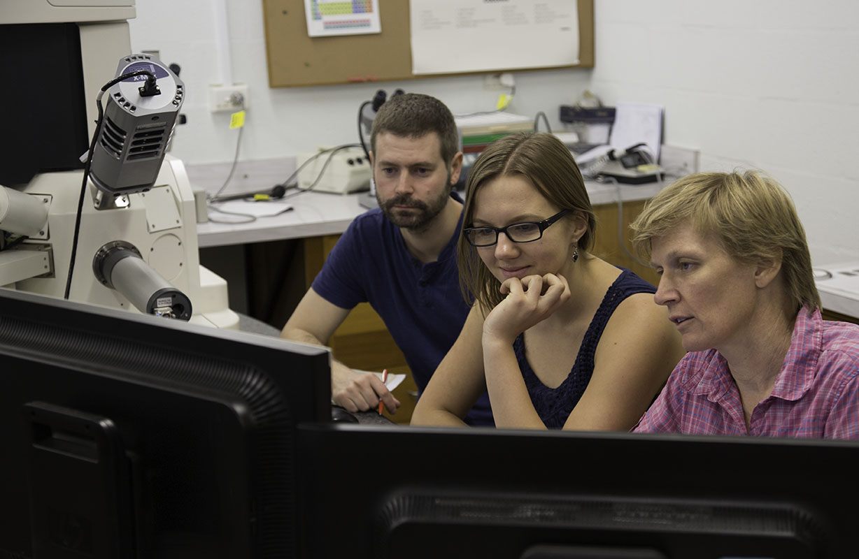
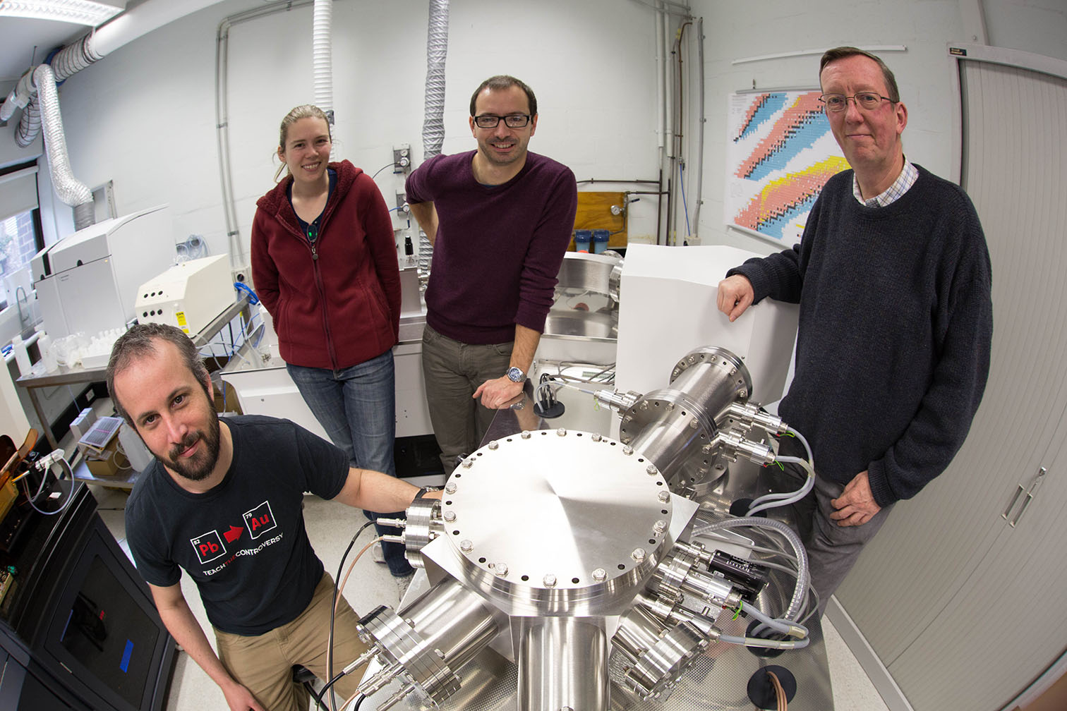
CCFS Visiting Researcher Dr Bjarne Almqvist, Daria Czaplinska and Sandra Piazolo using the SEM to examine advanced crystallographic orientation and simultaneously acquired chemical data from New Zealand lower crustal rocks.
a. Electron Microprobe: The Zeiss EVO MA15 SEM carried the electron imaging workload providing high-resolution BSE and CL images for TerraneChron®(http://www.gemoc.mq.edu.au/TerraneChron.html) and other research projects, including diamonds, corundum, PGM in chromitites and experimental petrology.The Oxford Instruments AZtec Synergy combined Energy Dispersive System and Electron Back-Scatter Diffraction detector installed in 2012 providessimultaneous elemental and crystal orientation mapping. This capability has enabled new research directions in the study of deformation processes in mantleand crustal rocks, including melt/rock interaction in high-grade metamorphic rocks and metasomatism of mantle-derived peridotites, pyroxenites andchromitites. The Cameca SX100 electron microprobe (fitted with 5 wavelength dispersive spectrometers and a Bruker Energy Dispersive Spectrometer system)continued to service the demands for quantitative mineral analyses and X-ray composition maps for all projects including analysis of corundum andinclusions from kimberlitic tuffs; analysis of base metal sulfides and platinum group minerals; minor and trace element analysis of metals.
b. Laser-ablation ICPMS microprobe (LAM): In 2015 the combination of the Photon Machines G2 laser system and Agilent 7700 ICP-MS was used for all in situ trace element analyses and U-Pbgeochronology. The facility was used by 15 Macquarie PhD thesis projects, 5 international visitors, 3 Masters Research students, 6 users from otherAustralian institutions and several in-house funded research projects and industry collaborations. Projects included the analysis of minerals frommantle-derived peridotites, pyroxenites and chromitites, high-grade metamorphic rocks and biominerals. U-Pb analysis of zircon was again a major activitywith geochronology projects (including TerraneChron® applications: http://www.gemoc.mq.edu.au/TerraneChron.html) from Australia (NSW, SA, WA), NewZealand, China, Cuba, Israel, Chile, Lao People’s Democratic Republic, Oman and Russia. Method development also continued for the U-Pb dating of baddelyiteand rutile.
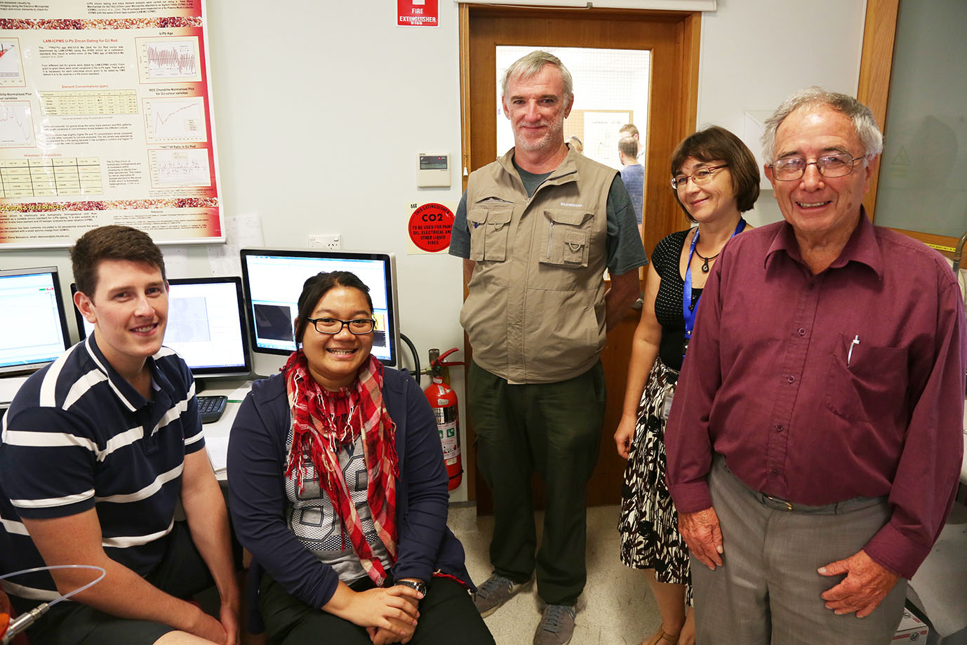
Chad Gardner, Angela Lay, Dr Ian Graham (School of BEES, UNSW) with Elena Belousova and Dr Lin Sutherland (Australian Museum & UWS) working on a collaborative project analysing trace-elements of gem stones (sapphire, ruby) and gem zircon from Australia and SE Asia.
c. MC-ICPMS:A Nu Plasma II MC-ICP-MS was installed in June 2015 and followed the decommissioning of Nu Plasma 005, after 16 years of service. Although the Nu Plasma IIrepresents a significant advance in its electronics and engineering, much of the fundamental design is adapted from Nu Plasma I. This has enabled arelatively seamless transition of existing methods developed over the past 15 years on the Nu Plasma I. The combination of the expanded collector array (16 Faraday cups and 5 ion counters) and enhanced sensitivity compared to the first generation Nu Plasma instruments has enabled the refinement of several in situ techniques pioneered at GEMOC, Macquarie.
The in situ measurement of U-Pb isotopes in zircon using the combination of the femtosecond laser system and Nu Plasma II is a world first, andpreliminary results were reported at the Goldschmidt Conference in Prague, August 2015 ( N.J. Pearson, W. J. Powell, Y. Gréau, R.C. Murphy, J.L. Payne, E. Belousova, W.L. Griffin and S. Y. O’Reilly 2015. U-Pb geochronology of zircon by femtosecond laser ablation. Goldschmidt Abstracts, 2015, 2437). The development of standard operating procedures for in situ U-Pb, Re-Os and Rb-Sr isotope measurements is on-going. The development of Mgisotope methodologies for chromite and chromite-rich ultramafic rocks as part of the TARDIS Program (Nicole McGowan PhD) in the ARC Centre of Excellencefor Core to Crust Fluid Systems (CCFS) was completed; high-precision results were obtained in wet-plasma mode on the Nu Plasma II and corroborated byreplicate measurements at IGGCAS, Beijing.
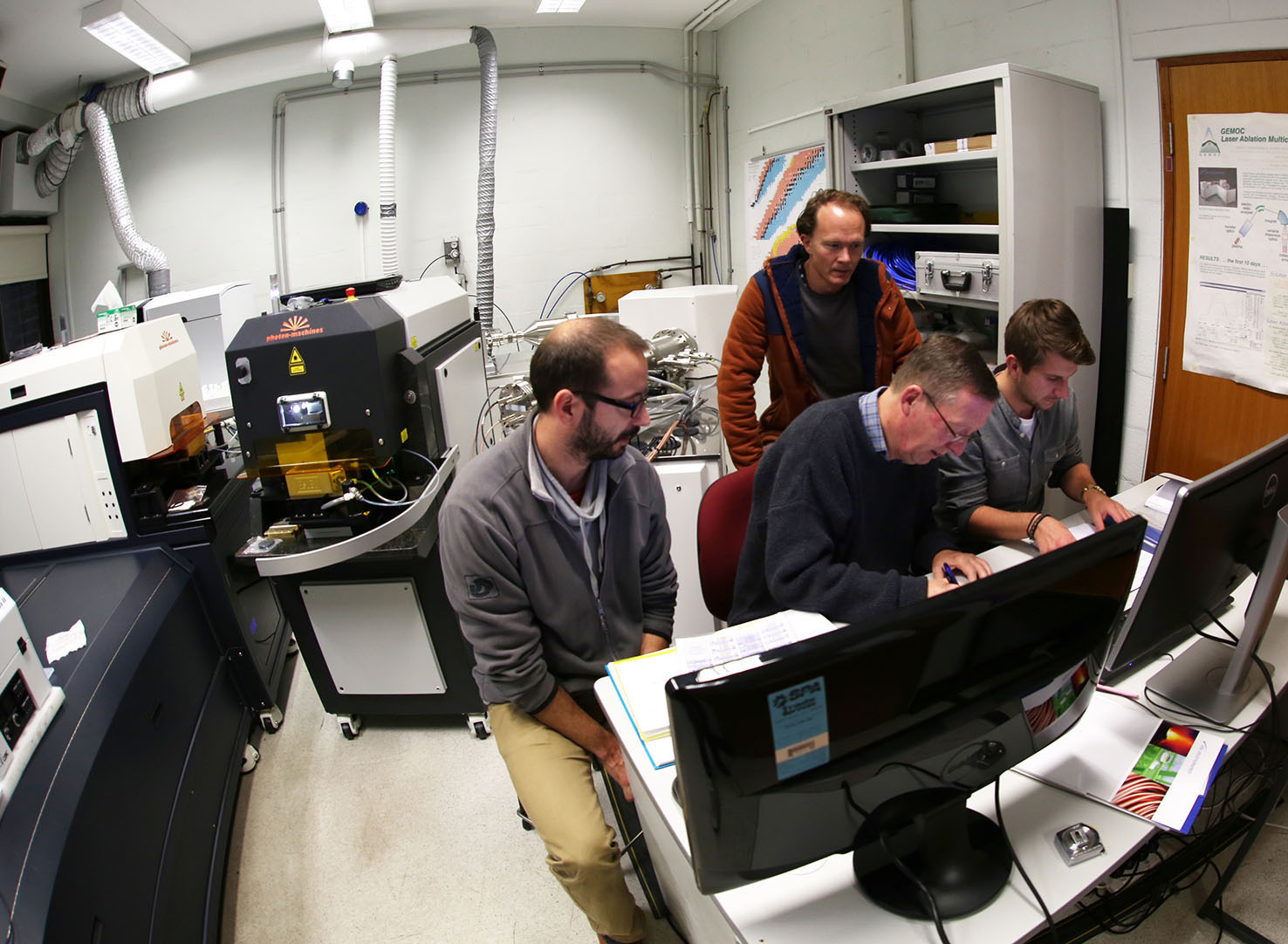
The sign off for the new Nu Plasma II MC-ICP-MS - Yoann Gréau, Will Powell, Norman Pearson and Nu instrument representative Jamie Royce.
At the time of the installation of the new Nu Plasma II, Nu Plasma HR 034 underwent an upgrade with an enhanced interface. The upgrade has increasedsensitivity between 1.5 and 2 times, and this has contributed to an overall improvement in signal stability, as well as in the precision of singlemeasurements and long-term reproducibility. In 2015 a third Photon Machines excimer laser microprobe was installed and co-located with Nu Plasma
HR 034.
The high demand for LAM MC-ICPMS time for in situ high-precision ratio measurements was again led by the analysis of Lu-Hf isotopes in zircon as amajor strand of the TerraneChron® activities (see http://www.gemoc.mq.edu.au/TerraneChron.html), with more than 1800 undertaken in 2015. In situHf isotopes were measured in zircons from Australia (NSW, WA, SA), Algeria, Bostwana, Brazil, China (Tibet), Indonesia, Iran, Israel, Laos, New Zealand,PNG and Russia. CCFS/GEMOC remains one of the few facilities with the capability to perform Re-Os dating of single grains of Fe-Ni sulfides and alloys inmantle-derived rocks. Re-Os studies were undertaken on xenoliths from eastern China, Siberia, Italy and Spain, and sulfide and platinum group minerals inchromitites from Tibet, Australia, Spain and Turkey.
GAU Director Norman Pearson (Right) acquainting the TerraneChron® Team, (L-R) Ed Saunders, Rosanna Murphy and Yoann Gréau, with the newly installed Nu Plasma II MC-ICP-MS.
d. Laboratory development: The clean-room facility established in 2004 continued to be used primarily for isotope separations for analysis on the Triton TIMS and Nu Plasma MC-ICPMS.Routine procedures have been established for Rb-Sr, Nd-Sm, Lu-Hf and Pb isotopes, as well as U-series methods (U, Th and Ra). In 2015 whole-rock Re-Osisotopic analysis of organic-rich sedimentary rocks was undertaken for the first time.
e. Software:GLITTER (GEMOC Laser ICPMS Total Trace Element Reduction) software is our on-line interactive program for quantitative trace element and isotopic analysisand features dynamically linked graphics and analysis tables. This package provides real-time interactive data reduction for LAM-ICPMS analysis, allowinginspection and evaluation of each result before the next analysis spot is chosen. GLITTER’s capabilities include the on-line reduction of U-Pb data. Salesof GLITTER are handled by AccessMQ and GEMOC provides customer service and technical backup. During 2015 a further 16 full licences of GLITTER were soldbringing the total number in use to more than 235 worldwide, predominantly in Earth sciences applications but with growing usage in forensics and materialsscience.
Dr Will Powell continued in his role in GLITTER technical support and software development through 2015. The current GLITTER release is version 4.4.4 andis currently available without charge to existing customers and accompanies all new orders.
2. X-Ray Fluorescence Analysis
In November 2012 a PANalytical Axios 1 kW X-ray Fluorescence Spectrometer was installed and is used routinely to measure whole-rock major elementcompositions on fused glass discs and trace-element concentrations on pressed-powder pellets. In 2013 the sample preparation equipment was upgraded andincluded a new furnace to make high-quality cast glass beads. The major element calibration was modified in 2015 to extend the spectrum of rock types thatcould be analysed to include Fe-rich samples such as iron ores and laterites.
3. Whole-rock solution analysis
An Agilent 7500cs ICPMS produces trace-element analyses of dissolved rock samples for the projects of CCFS/GEMOC researchers and students and externalusers, supplementing
the data from the XRF.
The ICPMS dedicated to solution analysis is also used to support the development of ‘non-traditional’ stable isotopes with the refinement ofseparation techniques and analytical protocols (see 1. d).
4. Diamond preparation and analysis
The GEMOC laser-cutting system (donated by Argyle diamonds in 2008) was used during 2015 to cut thin plates of single diamond crystals as part of theon-going research into diamond genesis. The plates are used for detailed spatial analysis of trace elements, isotopic ratios and the abundance andaggregation state of nitrogen. The nitrogen measurements are made using the ThermoFisher iN10 FTIR microscope, which allows the spatial mapping of wholediamond plates at high resolution with very short acquisition times using a software package developed here.
5. selFrag - a new approach to sample preparation
GEMOC’s selFrag instrument was installed in May 2010 and was the first unit in Australia. This instrument uses high-powered electrical pulses todisaggregate rocks and other materials along the grain boundaries. It removes the need to crush rocks for mineral separation, and provides a higherproportion of unbroken grains of trace minerals such as zircon. Since its installation selFrag has been used for a range of applications including zirconseparation, the analysis of grain size and shape in complex rocks, and the liberation of trace minerals from a range of mantle-derived and crustal rocks.
6. Spectroscopy
The spectroscopy infrastructure includes an FTIR microscope (ThermoFisher iN10 FTIR microscope; 2008). The FTIR is used to measure H abundance in a rangeof nominally anhydrous minerals (e.g. olivine, pyroxene, garnet) and H and N contents in diamond. In developing the spectroscopy capability an emphasis hasbeen placed on hyperspectral mapping to produce integrated datasets and multi-layered information in a spatial context. A Horiba H-CLUE CL monochromator(MQSIS 2015) is due to be installed on the Zeiss EVO SEM in January 2016. The monochromator system will provide imaging of individual emitting species andtheir distribution in a sample, and provide textural evidence of crystal growth, overgrowths and replacement, deformation, diagenesis and provenance.
Dr Christoph Lenz from ANSTO is our newest honorary associate using the Raman instrumentation.
7. Raman spectrometry
A confocal laser Raman microscope (co-funded by MQSIS 2014 and Future Fellowship funding to A/Prof Dorrit Jacob) delivers information for non-destructivephase-identification and characterisation at one micrometre spatial resolution. The Raman spectrometer continues to serve the CCFS, the Department and theFaculty. New acqusitions this year were a small Peltier cooled freezing stage, which is intended to help in the analysis of ephemeral phases in biomineralsbefore they re-crystallise. In addition, the facility is now extended to include a fibre probe head, which is planned to be used for in situ,realtime spectrometry of high pressure experiments. George Amulele and Simon Clark are working to design and build a sample platform for this purpose,which will open up new analytical avenues for the experimental petrology group at EPS. HDR student Sophia Bratenkov and MRes student Shirin Baydjanova fromthe organic geochemistry group have both discovered Raman spectrometry for the characterisation of organic compounds (kerogen) in geological samples.
8. Computer cluster
The cluster Enki has continued to be a powerhouse for the geodynamics group, supporting two funded research projects, 3 PhD projects, a postdoc, andnumerous Masters-level projects. Recent developments have included the development of planetary evolution capability of the mantle convection code Aspect(based on the deal.II finite element libraries), led by Siqi Zhang. In addition, O’Neill and Zhang have developed a new smoothed-particle hydrodynamic codeto simulate early solar-system processes, and have been utilising Enki for these simulations. A recent RIBG round to expand the lithosphere/seismic cluster“Toto” was successful (led by J.C. Afonso). In addition, a GPU Tower (supplied by Xenon systems) continues to act as a development machine for GPU-capablecode, including the recent SPH code, and a Xeon-Phi server (supplied by Dell) has recently been installed, enabling the modelling group to startdevelopment and migration of their codes onto this next generation hardware.
CMCA TECHNOLOGY DEVELOPMENT AND INSTRUMENTATION
The University of Western Australia’s Centre for Microscopy, Characterisation and Analysis (CMCA) is a $50M core facility providing analytical solutions across a diverse array of scientific research. The world-class facilities and associated technical and academic expertise are the focus of micro-analytical and characterisation activities within Western Australia, while strong links and collaborations have earned the CMCA an excellent national and international reputation. The CMCA incorporates the Western Australian Centre for Microscopy, and is a node of the NCRIS Characterisation capabilities, the National Imaging Facility (NIF) and the Australian Microscopy and Microanalysis Research Facility (AMMRF). It is also associated with the NCRIS funded Australian National Fabrication Facility (ANFF), and AuScope, which have made a substantial contribution to facilities run by CMCA.
CMCA capabilities:
- Secondary Ion Mass Spectrometry (CAMECA IMS 1280 and CAMECA NanoSIMS 50 and NanoSIMS 50L)
- Electron probe microanalysis (JEOL JXA 8530F)
- Focused ion beam (FEI Helios)
- Transmission electron microscopy (FEI Titan, JEOL 2100)
- Scanning electron microscopy (FEI Verios XHR, Zeiss 1555, Tescan Vega3)
- X-ray powder diffraction (Panalytical Empyrean)
- X-ray micro-CT (Xradia)
- Confocal Raman imaging with AFM (WiTec Alpha 300RA+)
- NMR spectroscopy (2 Bruker Avance and 2 Varian spectrometers)
- X-ray crystallography (Oxford Diffraction)
- GC and HPLC mass spectrometry
- Bioimaging, flow cytometry, cell sorting, and laser micro-dissection
- Optical and confocal microscopy
- Biological sample cryo-preparation and ultramicrotomy
THE AMMRF FLAGSHIP ION PROBE FACILITY
The CAMECA IMS 1280 and NanoSIMS 50 are flagship instruments of the AMMRF. The AMMRF Flagship Ion Probe Facility offers state-of-the-art secondary ion massspectrometry (SIMS) capabilities to the Australian and international research communities, allowing in situ, high-precision isotopic and elementalanalyses, and secondary ion imaging on a wide range of samples.
The IMS 1280 large-geometry ion probe, installed in 2009, was co-funded by the University, the State Government of Western Australia, and the Federal Government’s Department of Innovation, Industry, Science and Research (DIISR) under the ’Characterisation’ (AMMRF) and ’S tructure and Evolution of the Australian Continent’ (AuScope) capabilities of the National Collaborative Research Infrastructure Strategy (NCRIS).The NanoSIMS 50, installed in 2003, was funded through the Federal Government’s NCRIS-precursor, the Major National Research Facility scheme (NANO-MNRF).CMCA is also a part of the National Resources Science Precinct’s (NRSP) Advanced Resources Characterisation Facility (ARCF).
The Ion Probe Facility is a key characterisation component within the ARC Centre of Excellence for Core to Crust Fluid Systems.
To ensure the highest levels of quality and throughput, the CCFS has provided funding for a Research Associate position within the Ion Probe Facility, tofacilitate direct scientific and technical interaction for all CCFS users and projects.
The Ion Probe Facility is also a member of the International Atomic Energy Agency’s (IAEA) Network of Analytical Laboratories (NWAL), performing U isotopeanalyses on environmental samples for Nuclear Safeguards.
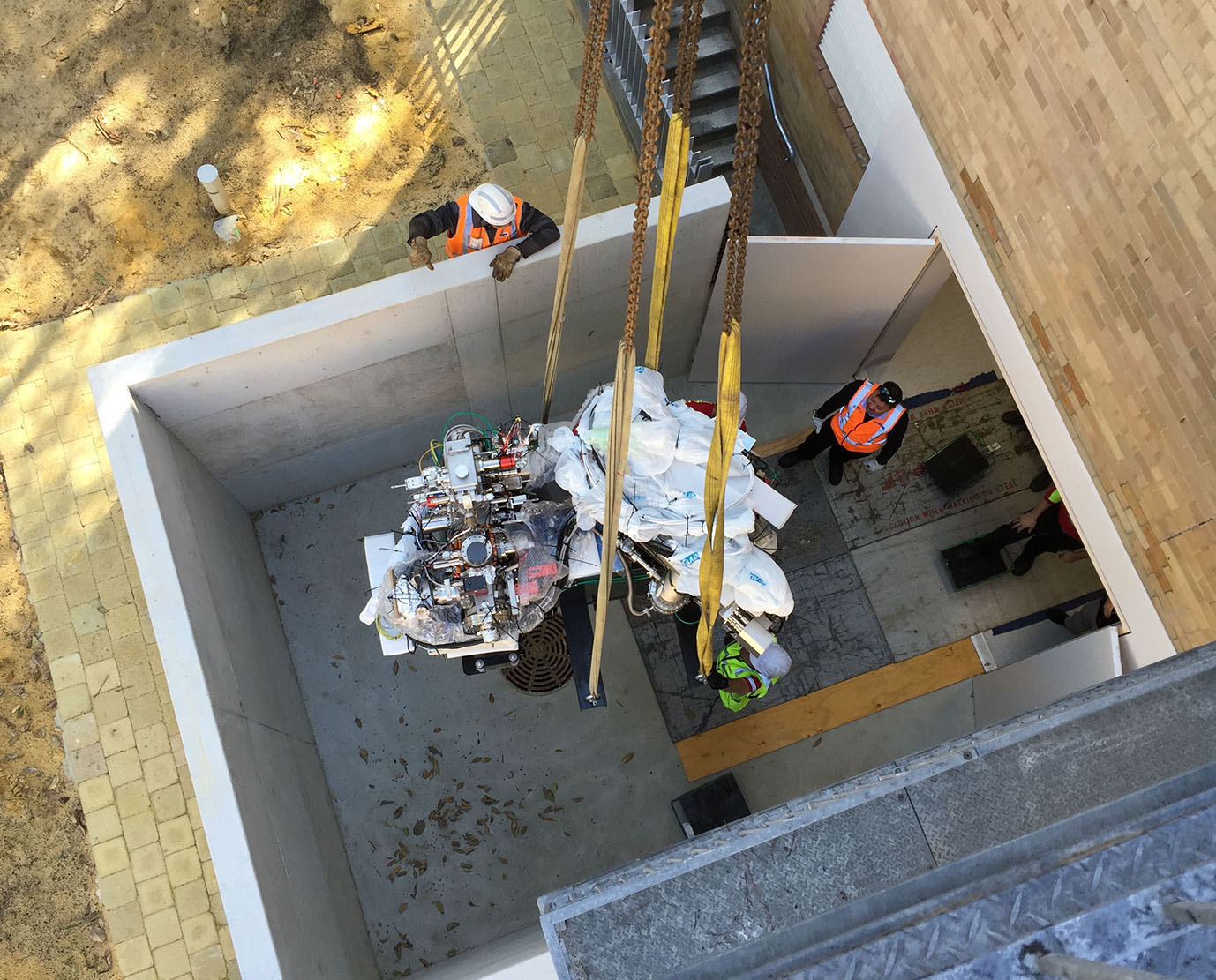
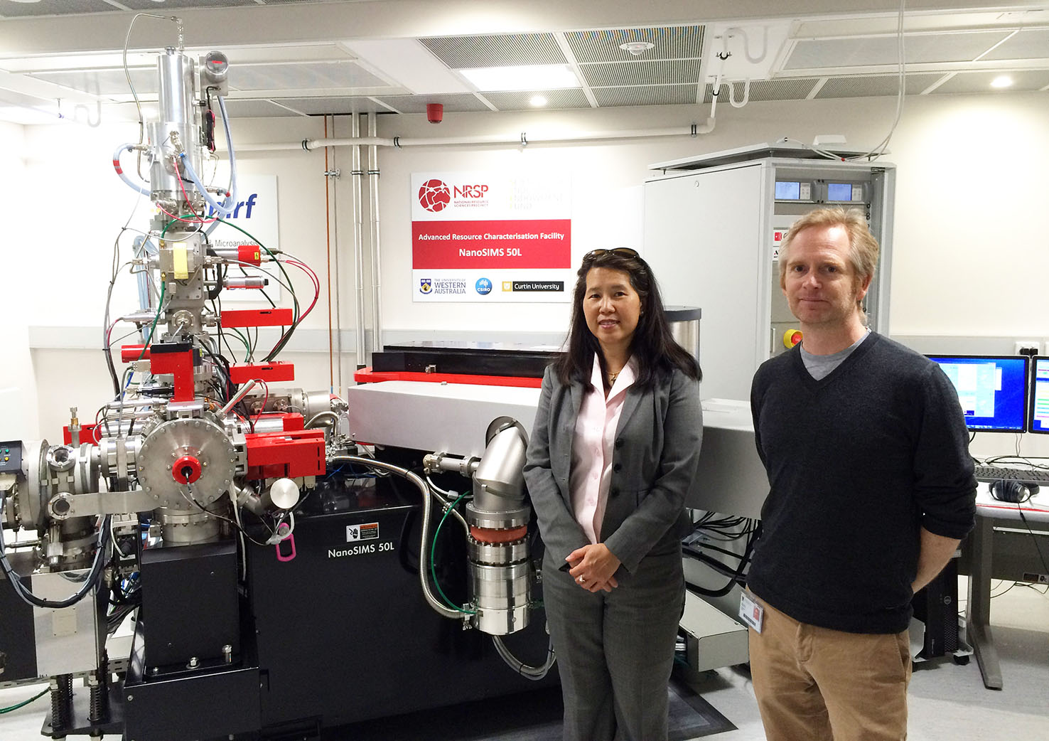
The CMCA’s NanoSIMS 50 being lowered into the new laboratory.
Progress IN 2015:
In 2015, the CMCA installed a new CAMECA NanoSIMS 50L ion probe as part of the NRSP’s Advanced Resources Characterisation Facility (ARCF). The ARCFprovides multiscale characterisation capabilities for Geoscience research, from the scale of drill core down to the atom scale. The Facility, fundedthrough CSIRO’s Science and Industry Endowment Fund (SIEF), features the Geoscience Atom Probe recently installed in the John de Laeter Centre at CurtinUniversity, and the synchrotron-less MAIA mapping facility under development at CSIRO. The NanoSIMS 50L represents a considerable technological advanceover the existing NanoSIMS 50, with a seven-FC/EM multicollector array and a new oxygen ion source allowing high-resolution isotope measurements ongeological samples. UWA contributed around $1M to build a new state-of-the-art laboratory to house both NanoSIMS instruments, and two new full-timepositions.
Recent successes in ARC LIEF funding have also updated the electron microscopy facilities at CMCA, with the installation in late 2015 of a FEI HeliosFocused ion beam (FIB) platform. The dual-beam instrument allows ion beam milling of samples for TEM, NanoSIMS and Atom Probe analysis, and high-resolution3D imaging.
2015 saw some personnel changes in the SIMS team at CMCA. Heejin Jeon joined the IMS1280 lab, and Haibo Jiang joined the NanoSIMS lab. Heejin is ageologist and SIMS expert with experience with both IMS1280 and SHRIMP. She completed her PhD at ANU, and then spent a 2-year research position at theNORDSIM facility in Stockholm (see her full profile on p.12). Haibo completed his PhD in materials science at the University of Oxford, developingNanoSIMS applications for a wide range of projects.
The Ion Probe Facility has continued to contribute to various projects in the context of CCFS. The IMS1280 clocked up almost 4000 hours, across more than20 projects. The majority of work involved multiple S isotope analyses with CET colleagues (Fiorentini, LaFlamme, Selveraja, Caruso), and O isotopes inzircon (Iaccheri, Bjorkman, Lu, Kirkland).
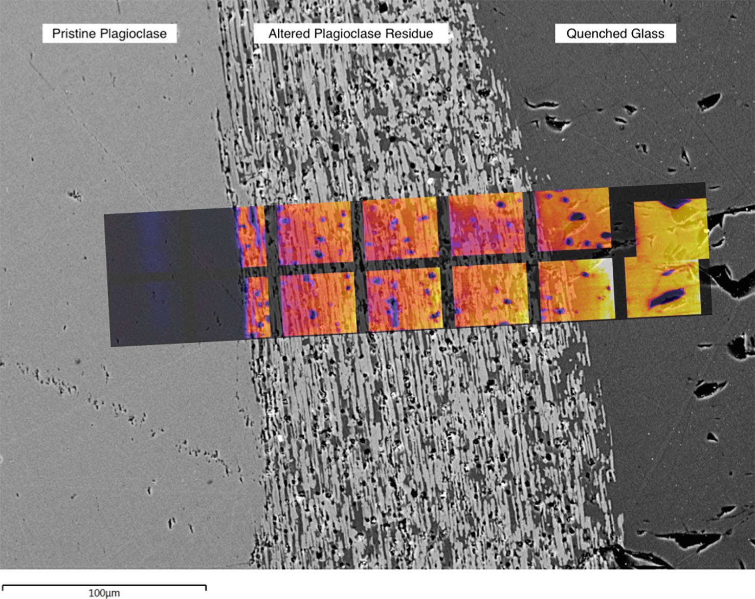
Isotope labels: The image shows the reaction interface between a plagioclase feldspar crystal on the left and hydrous silicate melt on the right. The SEM shows two sharp boundaries between the unreacted crystal, the reaction zone, and the melt. The overlain NanoSIMS image shows the oxygen isotopic composition across the reaction zone, and reveals that the reaction zone is enriched in 18O from the melt.
High-precision isotope measurement with SIMS requires calibration against known standards to correct for instrumental mass fractionation between analysissessions. This varies significantly between different materials, such that each new material analysed by SIMS necessitates the development of newstandards. 2015 saw a big push to develop in-house standards for a range of sulfide minerals - pyrite, pyrrhotite, pentlandite, chalcopyrite, andarsenopyrite. The results have been submitted for publication.
In 2015 the CMCA once again played an important role in a wide range of cross-nodal CCFS collaborative projects. The CCFS pilot project ' Making the Invisible Visible', involving the NanoSIMS to image isotope labels in experimental samples, began in earnest. UWA Honours students JackAdams and Haydn White visited Macquarie to perform experiments under the tutelage of Sandra Piazolo, Tracey Rushmer and John Adam, returning to UWA toperform the NanoSIMS analysis. Macquarie PhD student Liene Spruzeniece also visited the NanoSIMS lab to image experiments performed in Germany.
CMCA-CCFS 2015 publications: were published in high profile Journals such as Precambrian Research, Lithos, Geology Contributions to Mineralogy and Petrology and Earth and Planetary Science Letters: CCFS Publications #380a,489, 519, 527, 601, 742, 751, 771
CFS COO, Magdalene Wong Borgefjord, and Matt Kilburn viewing the newly installed CAMECA NanoSIMS 50L ion probe.
For further information on CMCA facilities please consult
http://www.cmca.uwa.edu.au/
JOHN DE LAETER CENTRE
The John de Laeter Centre (JdLC) is a collaborative research venture involving Curtin University, the University of Western Australia, CSIRO and theGeological Survey of Western Australia.
It hosts over $28M in infrastructure supporting research in: geosciences (geochronology, thermochronology and isotope studies); environmental science;isotope metrology; forensic science; economic geology (minerals and petroleum); marine science; and nuclear science.
The mission of the Centre is to “build world-class research infrastructure in Western Australia for the benefit of Earth, Environment and Materials Science research”. The JdLC isheadquartered in the Faculty of Science and Engineering at Curtin University, but has a governing board consisting of members of the joint venture partnersas well as representatives from the mining, petroleum and environment sectors.
The Centre experienced rapid growth in 2015 through the merger of mass spectrometry and microscopy facilities at Curtin, the commissioning of $5,200,000 innew analytical instrumentation and the appointment of 4 new research fellows. A new website has been developed to provide detailed information on the newfacilities, instrumentation and research staff (http://www.jdlc.edu.au).
The components of the JDLC are organised into fourteen major facilities:
A key milestone in 2015 was the establishment of the (GAP) Geoscience Atom Probe Facility as a node of the AdvancedResources Characterisation Facility (ARCF) funded by a $12,400,000 Science and Industry Endowment Fund grant to Curtin, UWA and CSIRO. The GAP hosts aCameca LEAP 4000X HR microscope (pictured below) capable of carrying out atom probe tomography (APT), a new characterisation technique combininghigh spatial resolution with time-of-flight mass spectrometry to provide 3-dimensional chemical information at the atomic scale. More commonly used tostudy semiconductors and metal alloys, the GAP is the first atom probe facility in the world to be dedicated to the study of geological materials(http://www.geoscienceatomprobe.org). The facility also commissioned a Tescan Lyra focused ion beam scanning electron microscope (FIB-SEM), with a Ga+ guncapable of micro-milling out a 100 nm wide needle of mineral sample prior to APT analysis. The Lyra system is a highly advanced platform for 2D and 3Dmicroanalysis with time of flight mass spectrometry (TOF-SIMS) and electron back scattered diffraction (EBSD) detectors. By correlating the analyticaloutputs of both the LEAP and the Lyra instruments, the ARCF provides an unprecedented capability of characterising highly complex materials on a wide rangeof length scales.
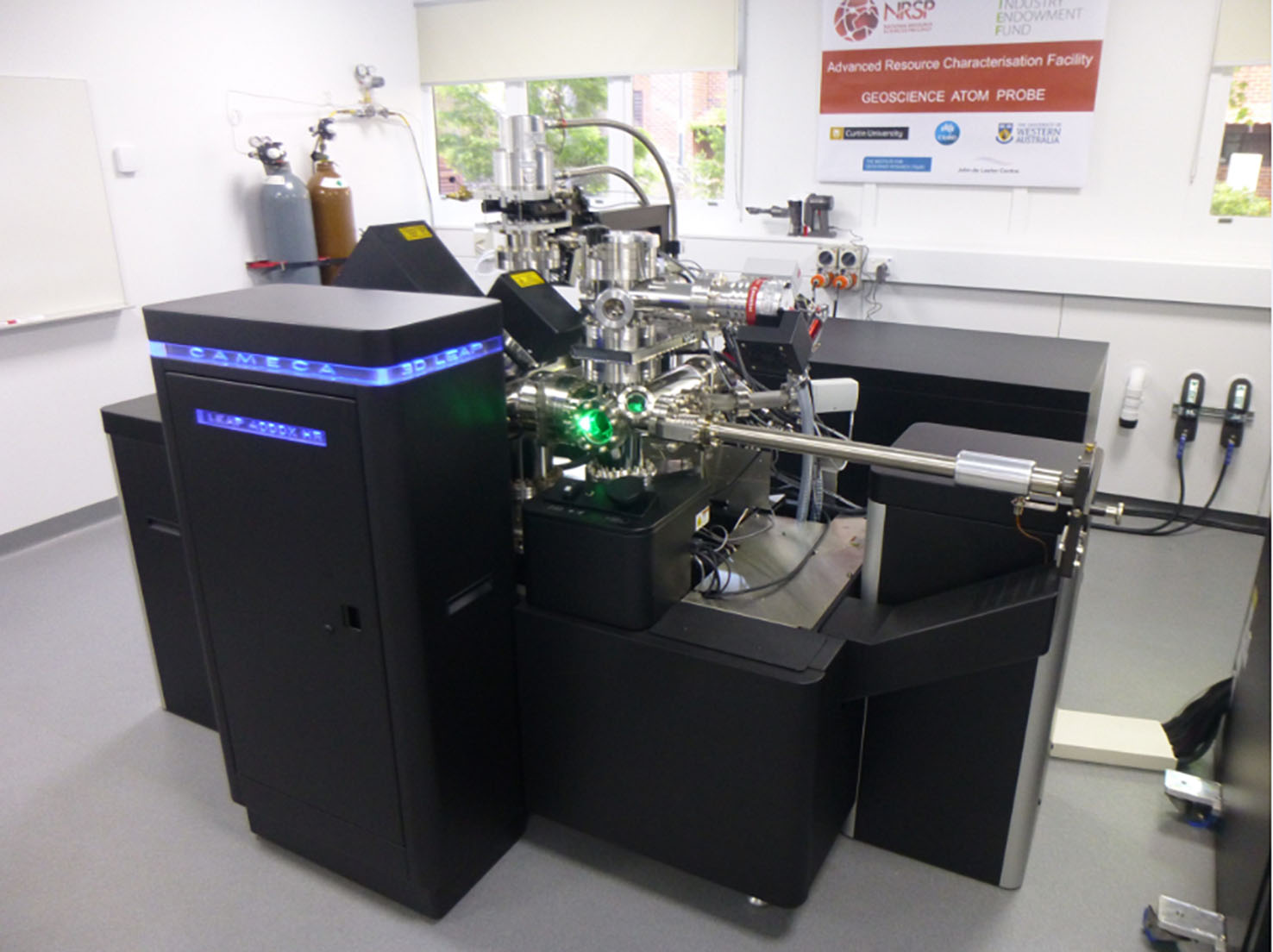
The Cameca LEAP 4000X HR microscope
The JdLC also commissioned a new (DMH) Digital Mineralogy Hub Facility with grant funding from the Australian Research Council supportedby Curtin University, University of Western Australia, Murdoch University and the Geological Survey of Western Australia. The Facility hosts a TescanIntegrated Mineral Analyzer (TIMA GM) - a fully automated, high throughput, analytical Field Emission Gun Scanning Electron Microscope (FEGSEM) forautomated analysis of sample composition. TIMA measures mineral abundance, liberation properties, mineral association and grain size automatically onmultiple samples of grain mounts, thin sections or polished sections. Applications include ore characterisation, process optimisation, remediation and thesearch for precious metals and strategic elements. The facility is being used by a broad spectrum of researchers: geologists and archeologists are usingthe facility in petrological characterisation, sample classification and lithofacies studies; while geochemists and geochronologists are using the mineralclassification outputs as targeting maps for further ion, electron or laser microprobe analysis. An automated mineralogy workshop held at Curtin inNovember 2015 attracted 25 participants from the local minerals and petroleum services industry.
(CEG) Curtin Experimental Geochemistry Facility:
CEG provides facility for experimental petrology, geochemistry and hydrogeochemistry at pressures and temperatures that range from those at the Earth’ssurface to those at the base of the Earth’s crust. The Facility contains:
• 2 x 150 ton end loaded piston cylinder presses
• Coretest hydrothermal apparatus
• Assorted furnaces to 1400 degrees C
• Assorted titanium and Teflon-lined bombs
(GHF) GeoHistory Facility:
The GHF houses state-of-the-art laser ablation inductively coupled plasma mass spectrometry (LA-ICPMS) equipment, in addition to a low temperaturethermochronology laboratory. The LA-ICPMS comprises a Resonetics S-155-LR 193nm excimer laser ablation system coupled to an Agilent 7700x quadrupole ICPMS. The Excimer laser is also coupled to a RESOchron helium analysis line for in situ (U-Th-Sm)/He, U-Pb and trace element analysis of single crystals . The facility also has a separate Alphachron helium line with a diode laser and furnace in order to facilitate conventional (U-Th)/He dating onsingle mineral crystals and larger samples. In late 2015 a Nu Plasma II multi-collector will be integrated into the facility to facilitate split streamanalysis.
(MMF) Microscopy and Microanalysis Facility:
The MMF houses a broad range of advanced microanalysis instrumentation providing high quality chemical, mineralogical and microstructural information, andhigh resolution images for research and technical publications. The facility staff have expertise in Materials and Earth Science research which is used tosupport both academic research and applied projects for the Western Australian minerals and energy sector. Techniques and instrumentation availableinclude:

• High resolution imaging (SEM, TEM) - The EVO is a variable pressure scanning electron microscope (VP-SEM). The microscope is suitable for general purposemicrostructural analysis at high vacuum, or for the analysis of non-conductive/hydrated samples at lower vacuum.
The JEM is a transmission electron microscope (TEM) with a LaB6 filament. The TEM is equipped with an EDS detector and a scanning TEM attachment. Thisinstrument is capable of elemental and microstructural analysis at extremely high magnifications.
• Spatially resolved elemental analysis (EDS) and Phase & orientation analysis (EBSD) - The MIRA3 is a variable pressure field emission scanningelectron microscope (VP-FESEM) that features sensitive EDS and EBSD detectors and integrated software for high quality microstructural analysis ofcrystalline samples.
• Quantitative mineral analysis (Q-XRD) - The D8A is an X-ray Diffractometer (XRD) with a copper x-ray source and an automated 45 position sample changer.It features a LynxEye position sensitive detector that is 200 times faster than a conventional scintillator detector, allowing collection of superior datain a short time-frame.
• Ion beam sample manipulation including TEM & TKD lamella preparation (FIB) - The NEON is a dual beam focused ion beam scanning electron microscope(FIB-SEM) equipped with a field emission gun and a liquid metal Ga+ ion source. This instrument combines high resolution imaging with precision ion beamablation of focused regions, allowing for site specific analysis of the surface and subsurface of samples in 2D or 3D.
The MMF also houses a suite of equipment that includes light microscopy, vacuum mount impregnation, manual and automated polishers, mills and coaters thatare used to prepare samples for electron microscopy and x-ray diffraction.
(SAXS) Small Angle X-Ray Scattering Facility:
The small angle x-ray scattering can be used to characterise the size, shape and distribution of objects between 1 and 100 nm. Instrumentation includes aBruker NANOSTAR SAXS comprising a copper sealed tube x-ray source with a gas filled two dimensional photon counting detector. 2015 LIEF funding will beused to upgrade the instrumentation in the facility.
(SHRIMP) Sensitive High Resolution Ion Micro Probe Facility:
The facility at Curtin has two automated SHRIMP II ion microprobes capable of 24-hour operation, together with a preparation laboratory that was remodelledin 2014. The equipment allows in situ isotopic analysis of chemically complex materials with a spatial resolution of 5-20 microns. The mainapplication of the SHRIMP instruments at Curtin is for U-Th-Pb geochronology of zircon and other U-bearing minerals, including monazite, xenotime,titanite, allanite, rutile, apatite, baddeleyite, cassiterite, perovskite and uraninite where multiple growth zones commonly require analyses with highspatial resolution. SHRIMP II is fitted with a Cs source, electron gun and 5 channel M/C. SHRIMP II A is currently being developed for stable isotopeanalysis of O in zircon and other silicates, and S in sulfides.
(SMS) selFrag & Mineral Separation Facility:
A selFrag facility, supported by an ARC LIEF grant, has been installed within the Department of Applied Geology at Curtin University. The facility provideselectric pulse disaggregation for mineral separation, which allows mineral grains to be separated from rock samples without the damage associated withstandard crushing techniques.
(TIMS) Thermal Ionisation Mass Spectrometry Facility:
The TIMS facility at Curtin incorporates a Thermo Finnegan Triton™ and a VG 354 multicollector mass spectrometer. The Triton is equipped with a21-sample turret and 9 faraday cups, enabling a precision of 0.001% on isotopic ratios. As well as geological applications within the broad field ofisotope geochemistry (Re/Os, U/ Pb, Pb/Pb, Sm/Nd, Rb/Sr) the TIMS instruments can be applied to a variety of isotope fingerprinting, such as forensics andthe environmental impact of human activities. The TIMS instruments are also used for the calibration of isotopic standards and the calculation of isotopicabundances and atomic weights. The facility has recently installed a Thermo Scientific Triton™ mass spectrometer, facilitating a new range ofgeochemical, geological and environmental research applications.
(TRACE) TRACE Research Advanced Clean Environment Facility:
This consists of a ~400m2 class 1000 containment space housing four class 10 ultra-clean laboratories, a class 10 reagent preparation laboratory
and a −18 °C class 10 cold clean laboratory, located at Curtin University. The extremely low ultimate particle counts are achieved with successive ‘spaces within spaces’ and HEPA filtration at each stage.
(WAAIF) Western Australian Argon Isotope Facility:
This is located at Curtin and is equipped with A MAP215-50 mass spectrometer with a low-blank automated extraction system coupled with a New Wave Nd-YAGdual IR (1064 nm) and UV (216 nm) laser, an electromultiplier detector and Niers source. The ultra-violet laser is capable of high-resolution (up to 10 µmbeam size) ablation of any mineral, allowing detailed analysis of individual mineral grains. The facility also houses an Argus VI Multi-Collector Noble GasMass Spectrometer.
The 40Ar/39Ar method is used to date a myriad of geological events such as volcanism, tectonic plate movements, mountain buildingrates, sediment formation, weathering and erosion, hydrothermal fluid movements, and alteration and diagenesis of minerals.
(WA-OIG) WA Organic and Isotope Geochemistry Facility:
WA-OIG is an internationally-recognised group contributing to world-class research in the fields of organic and stable isotope geochemistry; paleogenomicsand geomicrobiology. Available techniques are listed here: http://jdlc.edu.au/wa-organic-and-isotope-geochemistry-facility-wa-oig/
(WABC) The West Australian Biogeochemistry Centre:
The WABC is a centralised, stable isotope facility housed at UWA. The Centre brings together a critical mass of researchers in stable isotope science,particularly in its application to environmental and ecological problems in both terrestrial and marine environments. The following instrumentation isavailable:
• Automated Nitrogen Carbon Analyser with Isotope Ratio Mass Spectrometer
• SerCon Elemental Analyser (Gas Solid / Liquid Preparation Unit), coupled with:
• 20-22 Stable Isotope Ratio Mass Spectrometer (Sercon, Crewe UK)
• vPicarro Cavity Ring-down Spectrometer L1115-I
• Delta XL Isotope Ratio Mass Spectrometer (Thermo/Germany) connected with:
• GasBench II (GB)
• Thermal Conversion /Elemental Analyser (TC/EA)
• Delta V Plus Isotope Ratio Mass Spectrometer (Thermo /Germany) connected with:
• Thermo Elemental Analyser Flush 1112 via Conflo IV
• Finnigan LC-IsoLink
• Agilent 6890/5973B GC-MSD
(XSAF) X-Ray Surface Analysis Facility:
The Western Australian XSAF provides access to a state of the art X-ray photoelectron spectroscopy system for studying surface elemental and chemicalcomposition. X-ray photoelectron spectroscopy (XPS) is a non-destructive technique used to probe the elemental and chemical composition of the first 2-5 nmof a sample surface. All elements heavier than helium can be detected down to an atomic concentration of 0.1-1%. The primary instrument in the facility isthe Kratos AXIS Ultra DLD X-ray photoelectron spectroscopy system with imaging hemispherical analyser, monochromated Al and Ag X-ray sources, Ag/Mg X-rayflood source, argon gas cluster ion gun, field emission electron source and ultra-violet helium lamp.
For further information on JDLC facilities please consult
http://www.jdlc.edu.au
WESTERN AUSTRALIA PALEOMAGNETIC AND ROCK-MAGNETIC FACILITY
The Western Australia Paleomagnetic and Rock-magnetic Facility was established at the University of Western Australia by CCFS CI Z.X. Li in 1990, funded bya UWA start-up grant to the late Professor Chris Powell. It was subsequently upgraded through an ARC Large Instrument Grant in 1993 to purchase a thenstate-of-the-art 2G Enterprises AC-SQUID cryogenic magnetometer and ancillary demagnetisation and rock magnetic instruments. It was upgraded again in 2006with a 4k DC SQUID system plus a Variable Field Translation Balance (VFTB) through an ARC LIEF grant, into a regional facility jointly operated by CurtinUniversity, UWA and the Geological Survey of WA. A MFK-1FB kappa bridge was installed in 2011. In 2014, a national consortium consisting of CurtinUniversity, The University of Western Australia, the Australian National University, Macquarie University and University of Queensland was awarded an ARCLIEF grant to purchase a new 2G 755 superconducting rock magnetometer with a vertical Model 855 automated sample handler (the RAPID system) and otheraccessories (ovens etc.), to be housed in a purpose-built magnetically shielded room at Curtin University’s Bentley campus. The magnetically shielded roomwas constructed during July-August 2015 by Dr Gary Scot’s team, and the laboratory refurbishment is expected to be completed in February 2016. The newlypurchased AGICO JR-6A spinner magnetometer and a TD-48SC thermal demagnetiser are already serving the research group, and delivery of the 2G RAPID systemis expected in mid-2016. Dr Josh Beardmore has recently been appointed as the Technical Officer who will oversee the day-to-day operation of the facilityfrom 2016.
The joint WA facility is one of three similar laboratories in Australia. The new purchases will build on existing instruments in the facility, including:
- va 2G 755 superconducting rock magnetometer with a vertical Model 855 automated sample handler (the RAPID system) and other accessories
- 755 2G cryogenic magnetometer upgraded (LE0668377) to a 4K DC SQUID system (currently back to 2G for a minor upgrade and for repair of thelightning-damaged cold head)
- MMTD80, MMTD18 and TD-48-SC thermodemagnetisers
- Variable Field Translation Balance (VFTB)
- MFK-1FB kappabridge
- Bartington susceptibility meter MS2 with MS2W furnace
A wide range of research topics has been investigated using the facility, including reconstructing the configuration and drifting history of continents allover the world from the Precambrian to the present, analysing regional and local structures and deformation histories, dating sedimentary rocks andthermal/chemical (e.g. mineralisation) events, orienting rock cores from drill-holes, tracing ancient latitude changes, paleoclimates, and recentenvironmental pollution.
There are four major current research programs for the updated facility:
(1) Supercontinent studies
(2) Formation and evolution of the Australian continent
(3) Magnetostratigraphy and applications to petroleum
geology and palaeoclimatic studies
(4) Planetary science applications and the early Earth